SPONDYLOLISTHESIS
The term Spondylolisthesis describes the forward (anterolisthesis) or backward (posterolisthesis) slippage of one vertebrae in relation to the vertebrae below.
There are many subtypes of Spondylolisthesis, however, the most common types are isthmic and degenerative.
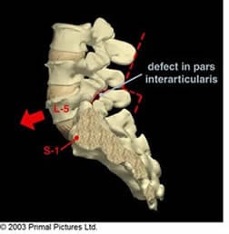
The Pars Interarticularis, or pars for short, is the part of vertebra located between the inferior and superior articular processes of the facet joint. It forms part of the ‘hook’ that enables the upper vertebra to remain in position thus playing an important role in intervertebral stability.
An Isthmic Spondylolisthesis commonly occurs between the lowest vertebrae (L5) and the sacrum (S1), whereby the Pars Interarticularis changes anatomically either secondary to a birth defect resulting in an intact but elongated pars,
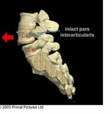
acute trauma or, more commonly, a stress fracture. With this latter spondy, repetitive hyperextension of the low back in young athletes, such as gymnasts, leads to the gradual development of a stress fracture and an interruption of the bony pars leading to what is termed a lytic spondylolisthesis.
A Degenerative Spondylolisthesis commonly occurs between the fourth and fifth lumbar vertebrae and presents in elderly women secondary to wear and tear at the discs and facet joints. Due to the degenerative changes within the discs and the joints at this level, patients often present with signs of nerve root irritation/compression due to stenosis.
SYMPTOMS
Symptoms arising from a Spondylolisthesis can vary from mild to severe with some patients having no symptoms at all, yet realising the presence of a Spondy purely as an incidental finding on x-ray.
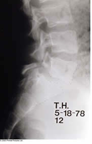
For other patients, symptoms and signs may include:
• Low back pain.
• Muscle tightness (hamstrings).
• Increased lumbar spine lordosis.
• Nerve root irritation/compression, i.e. sciatica.
Spondylolisthesis is often an incidental findings and only arises as a result of referring a patient for imaging. It is often seen in more athletic individuals, particularly those who were active during childhood and teen years. Typically, the low back pain is worse with extension and associated with an increased lumbar spine lordosis and tight hamstrings.
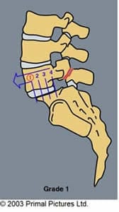
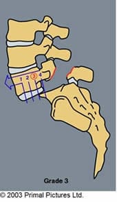
The radiographic grading of a Spondylolisthesis is based on dividing the sacrum (or L5) into fourths and observing the degree of slippage of the vertebrae above on the sacrum (or L5) below. Grade one and two are the most stable and can be treated conservatively, whereas the rarer grade 3 and 4 Spondylolisthesis are considered unstable and a surgical evaluation is necessary.
(The list of conditions given above and subsequent explanations are intended as a general guide and should not be considered a replacement for a full medical examination. Furthermore, we do not purport to treat all the conditions listed. Should you wish to discuss any of these conditions with our chiropractors, please do not hesitate to phone the clinic on 020 7374 2272 or email enquiries@body-motion.co.uk).
Our team of chiropractors and massage therapists are on hand to answer any questions you may have, so get in touch today via enquiries@body-motion.co.uk or on +44 (0)20 7374 2272.
